
Canine Leishmaniosis, often referred to as CanL, is a parasitic disease caused by the protozoan parasite Leishmania. This disease primarily affects dogs but can also affect other mammals, including humans. In this blog, we will delve into the various aspects of Canine Leishmaniosis, including its etiology, causes, symptoms, transmission, consequences, prevention measures, diagnosis methods, and treatment.
What is Canine Leishmaniosis?
Canine Leishmaniosis is a vector-borne disease caused by the Leishmania parasite, transmitted to dogs through the bite of infected sandflies. This parasitic infection can lead to a range of clinical manifestations, affecting various organs such as the skin, spleen, liver, and lymph nodes.
Various species of Leishmania are responsible for causing the disease, with Leishmania infantum being the primary culprit in many parts of the world. This parasite is endemic to the Mediterranean basin, South America, and Central and Southwest Asia, and is the most widespread Leishmania species.
Symptoms of Canine Leishmaniosis:
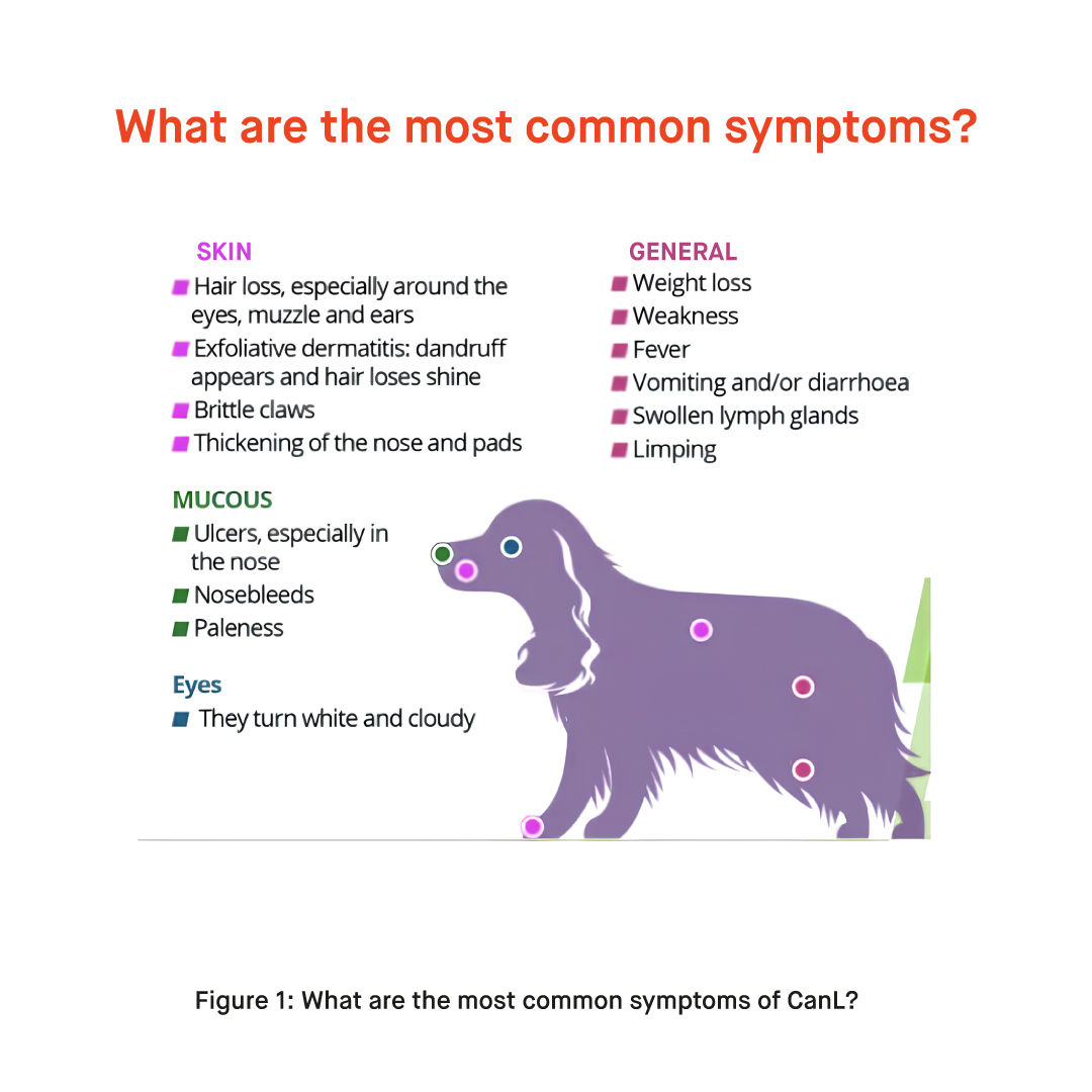
Canine Leishmaniosis (CanL) can manifest with a variety of symptoms, and the severity of the disease can vary from mild to severe. Symptoms include;
- Skin issues like nodules, ulcers, and hair loss around the eyes, ears, muzzle, and footpads.
- Systemic signs such as weight loss, lethargy, weakness, and fever may be observed. Swollen lymph nodes, especially around the neck and shoulders, are common.
- Ocular symptoms like conjunctivitis and uveitis can occur, along with joint pain, lameness, and arthritis.
- Renal symptoms may manifest as increased thirst and urination, while gastrointestinal issues like loss of appetite, vomiting, and diarrhea may also be present.
It’s important to note that not all dogs infected with Leishmania parasites will display symptoms. Some may carry the infection without apparent clinical signs, acting as asymptomatic carriers. Additionally, the progression and severity of symptoms can vary widely among individual dogs.
Transmission of CanL
Dogs can become infected with Canine Leishmaniosis (CanL) through the bite of an infected sandfly. Here’s how the transmission occurs:
1. Sandfly Transmission:
- Female sandflies are the primary vectors responsible for transmitting CanL.
- When an infected sandfly bites a dog during a blood meal, it injects the infective stage of the Leishmania parasite (promastigotes) into the dog’s bloodstream.
2. Vector-Borne Transmission:
- The Leishmania parasites then travel to various tissues within the dog’s body, where they transform into the amastigote stage and replicate.
- The amastigotes multiply within cells, particularly in organs like the spleen, liver, bone marrow, and skin.
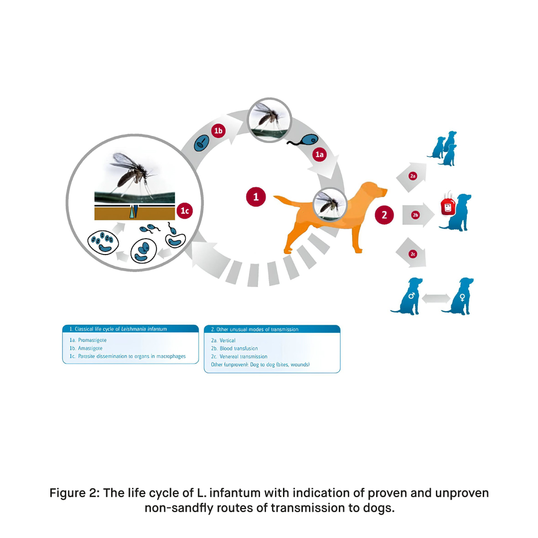
3. Vertical Transmission:
- In addition to vector-borne transmission, CanL can also be transmitted vertically from an infected mother to her puppies during birth or through the milk during lactation.
Prevention of CanL
Preventing CanL involves measures to reduce the risk of sandfly bites and managing the overall environment. Using insect repellents, insecticide-treated nets, and keeping dogs indoors during peak sandfly activity are effective preventive strategies.
Additionally, in regions where CanL is endemic, vaccination may be available to provide further protection against the disease. Regular veterinary check-ups and early intervention in case of symptoms are crucial for the well-being of dogs in areas where CanL is prevalent.
Treatment of Canine Leishmaniosis:
Treatment of Canine Leishmaniosis often involves a combination of medications, including antimonial drugs, allopurinol, and supportive therapies. Regular monitoring and follow-up care are crucial for managing the disease and preventing relapses.
Diagnosis Methods for Canine Leishmaniosis:
Diagnosing Canine Leishmaniosis involves a combination of clinical examination, laboratory tests, and specialized diagnostic methods. Here are the key diagnosis methods for CanL:
1. Clinical Examination: Thorough physical examination by a veterinarian to assess general health and observe visible symptoms, such as skin lesions, swollen lymph nodes, or ocular abnormalities.
2. Blood Tests:
- Serological Tests: Enzyme-linked immunosorbent Assay (ELISA) and Indirect Fluorescent Antibody Test (IFAT) to detect antibodies against Leishmania in the blood.
- Quantitative Polymerase Chain Reaction (qPCR): Molecular method to identify Leishmania DNA for a more specific diagnosis.
3. Bone Marrow Aspiration: Microscopic examination of bone marrow samples to detect Leishmania amastigotes within cells.
4. Cytology: Fine needle aspirates or tissue biopsies from affected organs, such as lymph nodes or skin lesions, examined microscopically for amastigotes.
5. Urinalysis: Examination of urine for signs of kidney involvement, a common effect of CanL.
6. X-rays and Imaging: Radiographic imaging to assess organ involvement, especially in cases of joint or skeletal abnormalities.
7. Eye Examination: Ophthalmic evaluation to detect ocular manifestations, including conjunctivitis and uveitis.
8. Lateral Flow Tests: Rapid diagnostic tests that use lateral flow technology for quick detection of Leishmania antibodies in blood samples.
A combination of these diagnostic methods, including the use of lateral flow tests, is often employed for accurate and timely detection of Canine Leishmaniosis. Early diagnosis is crucial for effective management and treatment. If a dog exhibits symptoms or resides in an area where CanL is prevalent, prompt veterinary attention and appropriate diagnostic tests are recommended for a comprehensive assessment.
How to Use Leishmania Rapid Test Kit in Dogs?
The Leishmania Rapid Antibody Test Kit is a diagnostic tool designed for the rapid and convenient detection of Leishmania antibodies in canine serum or plasma specimens. This test utilizes lateral flow technology to provide quick results. Here’s an overview of how it typically works:
Materials Provided with the Test Kit:
- Instruction Use
- Test Cassette
- Diluent
- Sample Dropper
- Sample Collection Tube (EDTA)
The procedure of Test:
Step 1: Sample Collection
- Clean the blood collection area with alcohol.
- Optionally use local anesthetic cream 20 minutes before blood collection.
- Draw enough blood directly from the vein using a syringe.
Step 2: Sample Processing
Serum:
- Collect whole blood into a non-anticoagulant tube, wait for coagulation, centrifuge, and collect the serum.
Plasma:
- Collect whole blood into an anticoagulation tube, centrifuge, and collect the plasma.
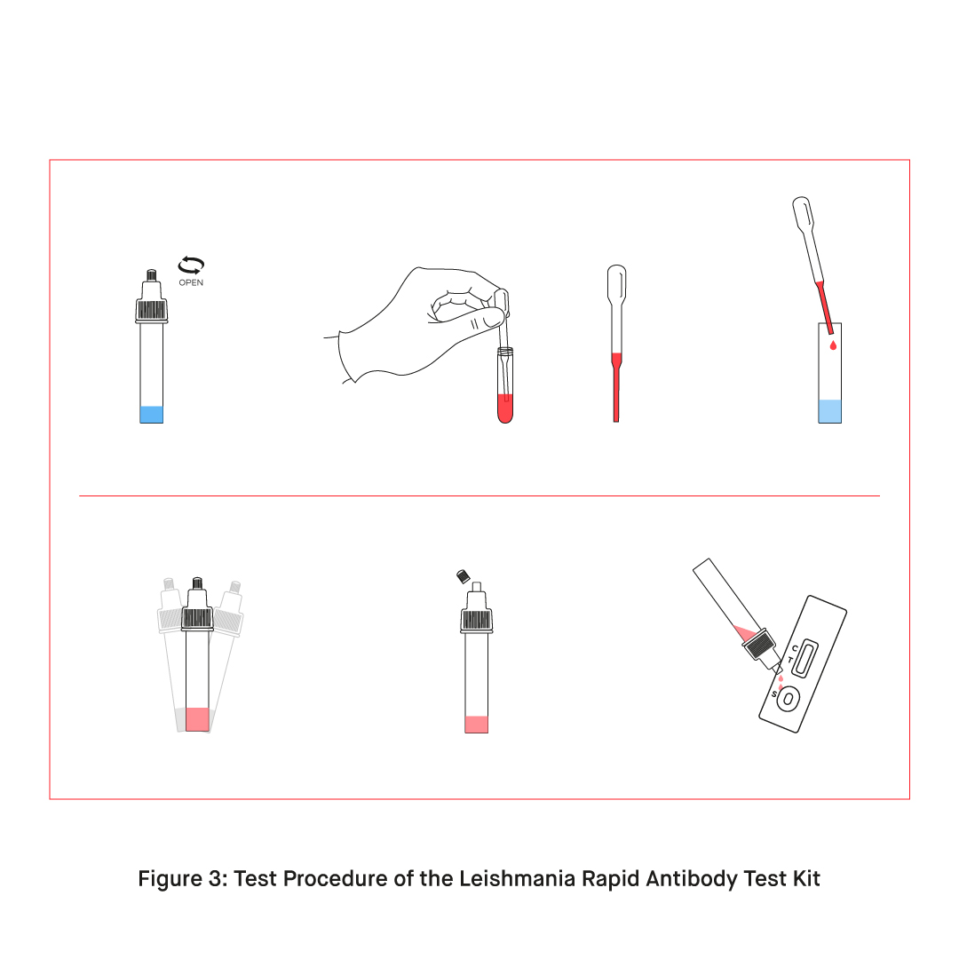
Step 3: Test Operation
- Open the test pack at room temperature and use it for a maximum of 15 minutes.
- Place the test cassette on a clean, flat surface.
- Open the tube cap.
- Using the capillary dropper, draw enough samples from the collection tubes.
- Add 20 µL of the sample to the tube containing the solution.
- Close the cap, mix, and carefully remove the release point on the tube cap.
- Add 3 drops of the sample-solution mixture to the sample well (S) on the cassette.
- Read the result after 10 minutes.
Step 4: Interpretation of the Result
- Positive Result: Both T and C lines appear.
- Negative Result: Only the C line appears.
- Invalid Result: The C line does not appear or only the T line appears; repeat the test with a new cassette.
Note: Do not read after 10 minutes.
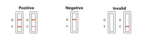
Figure 4: Interpretation of the results.
What are the Most Commonly Used Diagnostic Kits for Canine Leishmaniosis?
The Leishmania Rapid Antibody Test Kit is an advanced diagnostic tool designed for swift and accurate detection of Leishmania antibodies in canine serum or plasma. With a focus on speed, accuracy, and user-friendly operation, this kit facilitates early identification of Canine Leishmaniosis. Its rapid results and convenience make it a valuable asset for veterinarians and dog owners, enabling timely intervention and effective disease management in regions where Leishmania is prevalent.
The Leishmania Detection Kit employs Fluorescent RT-PCR for precise identification of Leishmania DNA in canine and feline samples. This advanced molecular diagnostic tool provides highly sensitive results, aiding in early detection and effective management of Leishmaniosis. The Fluorescent RT-PCR method enhances accuracy, making the kit invaluable for veterinarians and pet owners in regions where Leishmania is a concern for both canines and felines.
REFERENCESFormun Üstü
- Morales-Yuste, M., Martín-Sánchez, J., & Corpas-Lopez, V. (2022). Canine Leishmaniasis: Update on Epidemiology, Diagnosis, Treatment, and Prevention. Veterinary sciences, 9(8), 387. https://doi.org/10.3390/vetsci9080387
- Mahdavi, R., Martinkovic, F., Shams-Eldin, H., Pereira, I. E., Reis, A. B., Latz, A., … & Steinhoff, U. (2023). Comparative Study of a Novel Lateral Flow Rapid Test with Conventional Serological Test-Systems for the Diagnosis of Canine Leishmaniosis in Croatia and Brazil.
- Solano-Gallego, L., Miró, G., Koutinas, A., Cardoso, L., Pennisi, M. G., Ferrer, L., … & Baneth, G. (2011). LeishVet guidelines for the practical management of canine leishmaniosis. Parasites & vectors, 4, 1-16.
- Clinical staging, treatment, and prognosis. LeishVet. (n.d.). https://www.leishvet.org/fact-sheet/clinical-staging/